INTRODUCTION
Cervical cancer is a leading cause of morbidity and mortality among women worldwide, particularly in low- and middle-income countries. Early detection of precursor lesions through cytological screening, such as the Papanicolaou (Pap) test, has proven effective in reducing both the incidence of and mortality from this disease 1. Cervical squamous intraepithelial lesions are commonly categorized into three principal groups: atypical squamous cells of undetermined significance (ASC-US), low-grade squamous intraepithelial lesions (LSIL), and high-grade squamous intraepithelial lesions (HSIL) 2.
The Pap test, developed in the 1940s, has long been a cornerstone of cervical cancer screening 3. This procedure enables the identification of cellular changes suggestive of pre-cancerous or cancerous lesions. However, its performance may be influenced by factors such as specimen adequacy, personnel training, and adherence to regular screening intervals 4.
Despite advances in technology and screening approaches, reported detection rates for ASC-US, LSIL, and HSIL vary considerably across studies and populations. Such variability may reflect socioeconomic and cultural factors, as well as differences in access to health services 5. Therefore, a systematic review is warranted to characterize the prevalence of these lesions and to explore the factors associated with their detection.
This systematic review aims to analyze the existing scientific literature on the prevalence of ASC-US, LSIL, and HSIL detected by Pap testing, using a rigorous methodology based on the PRISMA protocol. Additionally, it aims to identify research trends and gaps that may inform improved strategies for cervical cancer prevention and early detection.
The overall aim of this study is to evaluate the prevalence of cervical intraepithelial lesions, including atypical cells and low- and high-grade lesions, through a bibliometric review based on Pap test findings. Specifically, the study will: (i) assess the prevalence of atypical cells across different populations; (ii) examine the frequency of low-grade intraepithelial lesions across regions and demographic groups; and (iii) evaluate the prevalence of high-grade intraepithelial lesions in diverse populations, thereby providing an integrated overview of these cervical abnormalities.
Literature Review
The prevention of cervical cancer has relied on timely detection of precursor lesions using cytological screening especially Pap test. Since its inception, the Pap test has been of significant importance in the prevention of incidence and mortality of cervical cancer by allowing the detection of cervical intraepithelial abnormalities before they could develop into invasive cancer. The traditional classification of cervical epithelial abnormalities as per the Bethesda system is based on (ASC-US), (LSIL), and (HSIL), each with its own biological behaviour, clinical and risk of malignancy 6.
The literature continuously underlines that early identification of cervical intraepithelial lesions is important to ensure the clinical intervention and effective management of the disease. ASC-US is the most common abnormal cytological result and often is transient cellular changes with the majority of such changes being linked to human papillomavirus (HPV) infection. Despite being classified as low-risk group, numerous studies have reported that some cases under this category may contain some underlying lesions of high grade, especially when there is the presence of high-risk HPV genotypes hence requiring stringent follow-up and triage measures 7.
The most typical association of LSIL is with transient HPV infections especially among the young women and has a high tendency of regression. However, chronic LSIL, particularly in combination with HPV high-risk types, can lead to higher-grade lesions, which can be emphasized by the necessity of proper surveillance and follow-up. Conversely, HSIL has a close relationship with chronic infection by oncolytic HPV genotypes and is an actual antecedent of invasive cervical cancer. Until diagnosed and treated, HSIL has a much greater potential to cause the development of cervical carcinoma, so its early detection should be part of the screening program.
The prevalence of ASC-US, LSIL, and HSIL in various populations and geographic areas has been explored in several studies recorded in large scientific databases such as Scopus, Web of Science, and Dimensions. The same studies have always documented significant inconsistency in the prevalence of lesions which has also been explained by variation in screening coverage, diagnostic standards, laboratory practice and population risk profile. HPV infection is reiterated to be the key etiological agent that leads to the pathogenesis and evolution of cervical intraepithelial lesions and sustained HPV infection with high risk is decisive in the progression of low-grade neoplasia to HSIL and invasive cancer 8.
The Pap test is the most commonly used screening tool in the world despite the introduction of HPV screening modalities especially in low- and middle-income nations as it is inexpensive, readily accessible, and has proven clinical applicability over time. Nonetheless, its diagnostic accuracy is affected by a number of conditions, such as the adequacy of the samples used, sampling methodology, the laboratory facilities, and also the training and experience of cytotechnologists and the pathologists 4. Fluctuation in these aspects may cause fluctuation in the detection rate and diagnostic accuracy which add to inconsistencies in the literature.
Besides technical factors, the socioeconomic factors and access to healthcare services are also significant determinants of cervical lesion prevalence. Research has shown the rate of cytological abnormalities is higher in the populations where there is less access to organized screening programmes, inadequate following up and where health literacy is less. On the other hand, those regions that have a well-developed screening systems and strong follow-ups are likely to record lower prevalence of high-grade lesions; this is because of early detection and proper treatment of precursor abnormalities 5.
Since the reported prevalence rates have great heterogeneity and the contextual issues of access to healthcare, screening practices, and HPV epidemiology, a systematic synthesis of current evidence is justified. A broad literature review and systematic review will help in understanding the prevalence trends of ASC-US, LSIL, and HSIL in the world in a better manner, gaps in the literature, and enable enhancement of cervical cancer screening approaches. This is necessary in order to communicate evidence-based public health policies that will help in reducing the disparities in cervical cancer prevention as well as enhancing the health outcome of women all over the world.
METHOD
Study Design
This systematic review was conducted in accordance with the Preferred Reporting Items for Systematic Reviews and Meta-Analyses (PRISMA) 2020 guidelines, which ensure transparency, reproducibility, and methodological rigour in evidence identification, study selection, and reporting 10. The study selection process is illustrated using a PRISMA 2020 flow diagram (Figure 1). The review protocol was not registered in PROSPERO, as the study involved a qualitative synthesis of published prevalence data without a meta-analysis; however, all eligibility criteria and methodological steps were predefined to minimize selection bias and enhance methodological transparency.
Eligibility Criteria
Predefined inclusion and exclusion criteria guided study selection. Original research articles published between 2000 and 2024 were eligible for inclusion if they reported cervical cytological findings obtained through Papanicolaou (Pap) smear screening. Eligible studies included those reporting the prevalence of atypical squamous cells of undetermined significance (ASC-US), atypical squamous cells—cannot exclude high-grade squamous intraepithelial lesion (ASC-H), low-grade squamous intraepithelial lesions (LSIL), and high-grade squamous intraepithelial lesions (HSIL), as defined by the Bethesda System.
Studies were included if they involved female participants of any age undergoing cervical cancer screening in population-based, opportunistic, or clinical screening settings. Only peer-reviewed articles published in English were considered.
Exclusion criteria comprised studies not related to cervical cytology or Pap smear–based screening, studies in which the primary outcomes were based on histopathological diagnosis or biopsy without corresponding cytological data, and studies with insufficient methodological quality or incomplete reporting of prevalence data. Case reports, conference abstracts, narrative or systematic reviews, editorials, letters to the editor, and non–peer-reviewed publications were also excluded.
Information Sources
An extensive literature search was conducted in several international databases to guarantee the wide coverage of the relevant studies. The databases were searched in Scopus, Web of Science (WOS), Dimensions and Google Scholar. The sources were chosen in order to obtain peer-reviewed literature of both clinical, epidemiological, and public health focus in the area of cervical cancer screening.
Search Strategy
The systematic search strategy was formulated in accordance with the research aim and major concepts, such as cervical intraepithelial lesions, cytological abnormalities, and Pap testing. The Boolean operators used to combine controlled vocabulary and free-text terms were AND and OR since they involve ASC-US, LSIL, HSIL, and Papanicolaou testing.
All the search strategies used in detail of each database are represented in Table 1, and the number of records found is indicated as well. Only original research papers that were published in peer-reviewed journals were searched.
Table 1.
Search Strategy and Databases.
|
Database |
Search equation |
Nro. of records |
|
Scopus |
("Evaluation of Atypical Cells" OR "Atypical Cells" OR "Atypia”) AND (“Low-Grade Lesions" OR "LSIL" OR "Low-Grade Squamous Intraepithelial Lesions”) AND (“High-Grade Lesions" OR "HSIL" OR "High-Grade Squamous Intraepithelial Lesions”) AND (“Papanicolaou Staining" OR "Pap Stain" OR "Papanicolaou Test”) AND ( LIMIT-TO ( DOCTYPE , "ar") |
|
|
|
205 |
|
|
WOS |
"cervical intraepithelial" AND (ASC-US OR ASC-H OR LSIL OR HSIL) AND (Papanicolaou OR Pap test) |
273 |
|
Dimensions |
"cervical intraepithelial" AND (ASC-US OR ASC-H OR LSIL OR HSIL) AND (Papanicolaou OR Pap test) |
500 |
|
Google Scholar |
("Evaluation of Atypical Cells" OR "Atypical Cells" OR "Atypia") AND (“Low-Grade Lesions" OR "LSIL" OR "Low-Grade Squamous Intraepithelial Lesions”) AND (“High-Grade Lesions" OR "HSIL" OR "High-Grade Squamous Intraepithelial Lesions”) AND (“Papanicolaou Staining" OR "Pap Stain" OR "Papanicolaou Test" ) |
|
|
|
190 |
Elaboration: The author.
Study Selection Process
The selection of the studies was done in a number of stages. The first step was search of records: in total 1,168 records were found in the four databases: 205 in Scopus, 273 in Web of Science, 500 in Dimensions, and 190 in Google Scholar.
Any records that had been retrieved were filtered in order to eliminate duplicates. The rest of the articles were then sifted by the title and abstract screening to determine relevancy to the research objectives. Assessment of potentially eligible studies was then done in full-text to ensure that the inclusion criteria are met. After this strict screening, 34 articles were found to be relevant and incorporated in the final analysis.
Data extraction and synthesis will be conducted on the data to draw conclusions about the designated study subject (Bruni et al., 2015).
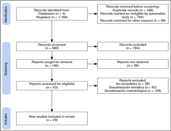
Figure 1. Prisma Flow Chart.
Elaboration: The author.
Data Extraction and Synthesis
The data will be extracted and synthesized to make conclusions regarding the predetermined study topic. Data were extracted into the relevant variables in each of the included studies, that is, study location, population, screening method, and prevalence of ASC-US, LSIL, and HSIL. Since the study designs, populations, and forms of reporting were diverse, the synthesis of the findings was done not by meta-analysis, however by a descriptive approach. Such a systematic synthesis allowed making meaningful comparisons of prevalence patterns in different geographic and healthcare settings.
Quality Assurance
To increase the credibility of the review findings, the studies underwent methodological rigour, relevance, and clarity of reporting evaluation stage at the full-text evaluation stage. Only studies of a satisfactory quality were used, so the synthesis of which can be considered a strong and reliable source of scientific knowledge.
RESULTS
Study Selection
After the systematic search and screening, 34 studies were identified to fit into the predefined inclusion criteria and were incorporated in the final qualitative synthesis. These researches documented the presence of atypical squamous cells (ASC-US), (LSIL), and (HSIL) using Papanicolaou (Pap) tests in different geographic locations and health facilities. It was noted that there was a certain level of heterogeneity in the prevalence rates reported which was a result of the differences in population factors and screening and diagnostic procedures.
Prevalence of Atypical Squamous Cells of Undetermined Significance (ASC-US)
There was a significant degree of variation between the 29 studies that reported the prevalence of ASC-US. This heterogeneity is seen to be determined by the variation in epidemiological context, screening equipment, and cytological interpretation.
A repeated study in Saudi Arabia by Abdallah et al. 11 and Ali-Risasi et al. 12 has reported an extremely high prevalence of ASC-US 40, which means that the burden of cytological abnormalities is high. This high rate can be explained by low screening uptake, the sufficiency of specimens, and inconsistency in the skills of cytotechnologists. Equally, the reports by Al Niyazee et al. 13 and Bonvicino et al. 14 in Iraq found a prevalence of 23.3% which, despite being lower as compared to that in Saudi Arabia, is still considered to be significantly high and may indicate inequities in access to healthcare services and delay in diagnosis.
Conversely, the ASC-US prevalence described by Barcelos et al. 15 in Brazil was significantly lower at 4.98 percent, which was in agreement with the range of 2 to 7 percent expected by the Bethesda System (2008). This observation could be attributed to a higher percentage of screening cover and more efficient follow-up programs, which allows the management and detection of abnormalities in the cervix at an earlier stage.
Also a number of studies explored the clinical implications of ASC-US. Barron et al. 16 and Çetiner et al. 17 proved that ASC-US can provide a significant risk of high-grade cervical intraepithelial neoplasia (CIN2/3), even to HPV-negative women. Such results indicate that further monitoring of ASC-US cases regardless of HPV status is important. Moreover, Bruno et al. 18 and Dhaubhadel et al. 19 found that the presence of HPV E6/E7 mRNA among women with ASC-US is linked to the risk of progression which is significantly high meaning that it could be used as a molecular marker in risk stratification. The results of Demarco et al. 20 and Dursun et al. 21 also supported the idea that a combination of ASC-US and high-risk HPV positivity is closely related to the development of CIN2+, which is why closer follow-ups in this subgroup are necessary.
Prevalence of Low-Grade Squamous Intraepithelial Lesions (LSIL)
The issue of LSIL prevalence used in the included studies also differed across the studies. In spite of the fact that LSIL usually carries lower malignant potential than HSIL, the prevalence and persistence of LSIL are still clinically significant, especially in areas with a lack of screening and follow-up.
Al Niyazee et al. 13 and Fonseca et al. 22 have reported exceptionally high levels of LSIL prevalence in Iraq (48%). The observation can be a result of the lack of screening programmes and the sexual health education, and increased prevalence of persistent HPV infection. Conversely, Indian based studies by Bal et al. 23 and Hirachand et al. 24 seem to reveal an LSIL prevalence of 2.7, which is in agreement with areas where more formalized screening programmes are established.
The close correlation between HPV infection and LSIL was also constantly true. Hong et al. 25 and Papestiev et al. 26 in North Macedonia found 46.7% of women were infected with HPV among patients with LSIL. The study conducted by De Brot et al. 27 also established that several high risk HPV infections have a strong relationship with LSIL persistence, which serves to highlight the need to follow up. Poomtavorn et al. 28 found LSIL prevalence in HPV positive women in Thailand to be 5.5 per cent which was similar to Saad et al. 29 in an urban US population. Collectively, these researches demonstrate the worldwide incidence of LSIL in various healthcare facilities.
In Kenya, there was no literature that reported exact LSIL prevalence rates, however, as Huertas et al. 30 and Muitta et al. 31 pointed to the importance of regular screening and early management in preventing the evolution of a lesion, in particular, in resource-limited settings.
Prevalence of High-Grade Squamous Intraepithelial Lesions (HSIL)
HSIL is the most shocking clinical precursor to cervical invasive cancer. Within the studies incorporated, HSIL prevalence was characterized mostly by a high level of variability, which depended on the availability of screening, the quality of follow-up, and population risk profiles.
Al Niyazee et al. 13 revealed that the HSIL prevalence in Iraq is 29, which is an extreme disease burden probably contributed by insufficient preventive screening and late diagnosis. On the other hand, a much lower prevalence of HSIL was reported by Abdallah et al. 11 at 0.55% in Saudi Arabia, possibly due to disparities in access to healthcare, epidemiology of HPV, or screening.
According to Cohen et al. 32 in the United States, a 1.6% progression rate of cytological category closely resembling HSIL was observed in women with ASC-H cytology, which supported the significance of high-follow up in cytological groups. The study by Sherman et al. 33 on the ALTS highlighted the need of active surveillance and early treatment of HSIL in order to avoid the development of invasive cancer. Further testimony of Rwanda by Shiramba et al. 34 also indicated a strong increase in the progression of untreated cases of HSIL, which indicated problems associated with low-resource conditions. Lastly, Syler et al. 35 emphasized the benefit of high-risk HPV genotyping in streamlining HSIL management by distinguishing women who were at highest risk of evolution, which can be used to implement a more focused and effective intervention approach.
DISCUSSION
This systematic review demonstrates substantial variability in the prevalence of atypical squamous cells of undetermined significance (ASC-US) and low- and high- grade squamous intraepithelial lesions (LSIL and HSIL) detected by the Pap test. These findings reflect important differences in epidemiological contexts, screening practices, and cytological interpretation across regions worldwide.
Interpretation of findings
In Saudi Arabia, Abdallah et al. 11 and Ali-Risasi et al. 12 reported an ASC-US prevalence of 40%, an alarmingly high figure indicating a considerable burden of cytological abnormalities. This may be attributable to limited screening coverage, specimen quality issues, and variability in cytotechnologist expertise. In Iraq, Al Niyazee et al. 13 and Bonvicino et al. 14 reported a prevalence of 23.3%, which remains high and may be influenced by unequal access to healthcare and delayed detection. Conversely, Barcelos et al. 15 in Brazil reported an ASC-US prevalence of 4.98%, consistent with the Bethesda System’s expected range (2–7%). This lower estimate may reflect stronger screening infrastructure and follow-up systems, support earlier detection and reduce the proportion of advanced disease.
Regarding ASC-US progression, Barron et al. 16 and Çetiner et al. 17 found that ASC-US may still pose a significant risk of progression to high-grade lesions (CIN2/3) even in HPV-negative women. This underscores the need for careful follow-up irrespective of HPV status. Moreover, Bruno et al. 18 and Dhaubhadel et al. 19 showed that E6/E7 mRNA detection is associated with a substantially higher risk of progression, suggesting its potential role as a clinically useful marker for risk stratification and more tailored management. Finally, Demarco et al. 20 and Dursun et al. 21 highlighted that ASC-US combined with HPV positivity is strongly associated with progression to CIN2+, supporting more intensive surveillance for this subgroup. With respect to LSIL, the reported prevalence ranged widely. Al Niyazee et al. 13 reported an LSIL prevalence of 48% in Iraq exceptionally high relative to other settings potentially reflecting limited screening programmes and insufficient sexual health education, leading to higher HPV incidence and LSIL detection. In contrast, Bal et al. 23 in India reported an LSIL prevalence of 2.7%, more consistent with organised health systems and established screening programmes. Papestiev et al. 26 identified HPV infection in 46.7% of women with LSIL in North Macedonia, supporting the strong association between HPV and LSIL. De Brot et al. 27 further noted that multiple high-risk HPV infections are significantly linked to LSIL persistence, emphasising the importance of continued surveillance. Similarly, Poomtavorn et al. 28 in Thailand reported an LSIL prevalence of 5.5% among HPV-positive women, indicating that LSIL remains clinically relevant even in settings with adequate access to screening, consistent with Saad et al. 29 in the United States. In Kenya, Muitta et al. 31 stressed the importance of regular screening and appropriate management to prevent progression to cervical cancer, particularly in resource- constrained environments.
For HSIL, Al Niyazee et al. 13 reported a prevalence of 29% in Iraq, highlighting a severe burden likely aggravated by limited preventive screening. In contrast, Abdallah et al. 11 reported an HSIL prevalence of 0.55% in Saudi Arabia, potentially reflecting differences in screening performance and population risk profiles. Cohen et al. 32 found that women with ASC-H cytology had a 1.6% progression rate to CIN2/3, underlining the need for rigorous management. Sherman et al. 33 (ALTS study) emphasised active surveillance in HSIL given the risk of progression to invasive cancer when not appropriately treated. Shiramba et al. 34 in Rwanda reported significantly higher progression among untreated HSIL cases, reinforcing the urgency of early, effective interventions. Syler et al. 35 demonstrated that high-risk HPV genotyping can optimise HSIL management by identifying women at greatest risk of progression, supporting more personalised strategies 36.
Practical implications
These findings have important implications for clinical practice and public health policy. The high prevalence of ASC-US and LSIL in settings with limited screening underscores the need to implement and strengthen cervical screening programmes to facilitate early detection and timely treatment. In addition, incorporating molecular markers such as E6/E7 mRNA testing in women with ASC-US may enhance risk stratification and improve clinical decision-making, potentially reducing progression to more severe disease.
Limitations
Several limitations should be considered. Heterogeneity in screening methods and cytological interpretation across included studies may have influenced prevalence estimates. Study quality also varied, with potential selection and information biases. Generalisability may be limited by differences in healthcare systems and demographic characteristics across populations.
Future directions
Future research should prioritise standardisation of screening approaches and cytological interpretation to support more accurate comparisons across regions. It is also essential to evaluate the impact of targeted interventions—such as sexual health education programmes and enhanced training for cytotechnologists—on reducing ASC-US and LSIL prevalence. Further studies on molecular markers and their clinical utility for risk stratification may substantially improve the management of cervical lesions.
CONCLUSIONS
This systematic review identified and evaluated the prevalence of cervical intraepithelial lesions, specifically atypical cells, low-grade lesions (LSIL), and high-grade lesions (HSIL) based on Pap test findings. First, ASC-US prevalence showed considerable variability, influenced by factors such as access to screening programmes and differences in cytological interpretation. This highlights the importance of robust health systems that ensure equitable access to high-quality screening services, thereby improving early detection and reducing observed variability. Second, LSIL prevalence likewise varied substantially across geographic regions and healthcare contexts. The findings demonstrate a strong correlation between HPV infection and the development of LSIL, reinforcing the need for HPV vaccination and sexual health education to reduce lesion incidence in vulnerable populations. Continued follow-up for women diagnosed with LSIL is also essential to prevent progression to more severe disease. Finally, HSIL prevalence represents a critical indicator of risk for the development of invasive cervical cancer. The variability observed across studies reflects disparities in screening access and follow-up quality. These findings underscore the urgency of strengthening early detection programmes and ensuring appropriate management of women with HSIL, particularly in resource-limited settings, to reduce the burden of cervical cancer. Overall, this review highlights the need for more comprehensive and accessible public health policies that address inequalities in the detection and management of cervical intraepithelial lesions. Integrating preventive strategies such as HPV vaccination and improved screening programmes is essential to reduce lesion prevalence and enhance women’s health outcomes globally.
Recommendations
Strengthen screening programmes: Implement and expand cervical screening in regions with limited early detection to ensure all women have access to regular Pap testing.
Education and awareness: Develop sexual health and HPV prevention campaigns to increase awareness of screening and vaccination benefits.
Training of healthcare personnel: Enhance training for cytotechnologists and healthcare professionals to improve accuracy in Pap test interpretation and reduce diagnostic variability.
Integration of molecular markers: Incorporate molecular testing (e.g., E6/E7 mRNA) to improve risk stratification in women with ASC-US and support optimized clinical decision- making.
Future research: Conduct further studies to standardize screening and cytological assessment methods, enabling more precise comparisons across regions and contexts.
The value of this review lies in its contribution to global understanding of cervical intraepithelial lesion prevalence and the disparities affecting their detection and management. By highlighting the need for stronger and more equitable health systems, this study provides evidence to support effective public health policies aimed at reducing cervical cancer incidence and improving women’s reproductive health worldwide.
CONFLICT OF INTEREST
The author declare that they have no conflict of interest in the publication of this article.
FINANCING
Non financing.
ACKNOWLEDGMENTS
To all the social agents involved in the development of the research.
REFERENCES CONSULTED
1. Organización Mundial de la Salud. Estrategia mundial para la eliminación del cáncer cervicouterino | UICC. [citado 14 de diciembre de 2024]; Disponible en: https://n9.cl/t5pej
2. Solomon D, Davey D, Kurman R, Moriarty A, O’Connor D, Prey M, et al. The 2001 Bethesda System: terminology for reporting results of cervical cytology. JAMA. 24 de abril de 2002;287(16):2114-9.
3. Sankaranarayanan R, Budukh AM, Rajkumar R. Effective screening programmes for cervical cancer in low- and middle-income developing countries. Bull World Health Organ. 2001;79(10):954-62.
4. Nayar R, Wilbur DC. The Pap Test and Bethesda 2014: “The reports of my demise have been greatly exaggerated.” (after a quotation from Mark Twain). Acta Cytol [Internet]. 19 de mayo de 2015 [citado 14 de diciembre de 2024];59(2):121-32. Disponible en: https://doi.org/10.1159/000381842
5. Yusheng Zhu, Sarah Feldman, Shuk On Annie Leung. Cervical Cancer Detection. 2023 [Internet]. 6 de marzo de 2023 [citado 14 de diciembre de 2024]; Disponible en: https://acortar.link/5kDeGc
6. Massad LS, Einstein MH, Huh WK, Katki HA, Kinney WK, Schiffman M, et al. 2012 updated consensus guidelines for the management of abnormal cervical cancer screening tests and cancer precursors. J Low Genit Tract Dis. abril de 2013;17(5 Suppl 1): S1-27.
7. Demarco M, Hyun N, Carter-Pokras O, Raine-Bennett TR, Cheung L, Chen X, et al. A study of type-specific HPV natural history and implications for contemporary cervical cancer screening programs. eClinicalMedicine [Internet]. 1 de mayo de 2020 [citado 14 de diciembre de 2024];22. Disponible en: https://acortar.link/I6rxjk
8. Castle PE, Stoler MH, Wright TC, Sharma A, Wright TL, Behrens CM. Performance of carcinogenic human papillomavirus (HPV) testing and HPV16 or HPV18 genotyping for cervical cancer screening of women aged 25 years and older: a subanalysis of the ATHENA study. Lancet Oncol [Internet]. 1 de septiembre de 2011 [citado 14 de diciembre de 2024];12(9):880-90. Disponible en: https://acortar.link/R0ytCL
9. Rodríguez AC, Schiffman M, Herrero R, Hildesheim A, Bratti C, Sherman ME, et al. Longitudinal study of human papillomavirus persistence and cervical intraepithelial neoplasia grade 2/3: critical role of duration of infection. J Natl Cancer Inst. 3 de marzo de 2010;102(5):315-24.
10. Moher D, Liberati A, M. Tetzlaff J. (PDF) Moher D, Liberati A, Tetzlaff J, Altman DG, Group PPreferred reporting items for systematic reviews and meta-analyses: the PRISMA statement. PLoS Med 6: e1000097. ResearchGate [Internet]. 21 de noviembre de 2024 [citado 14 de diciembre de 2024]; Disponible en: https://acortar.link/Y5XSI
11. From a large referral hospital in Saudi Arabia using the revised 2001 Bethesda System [Internet]. [citado 15 de diciembre de 2024]. Disponible en: https://www.annsaudimed.net/doi/epdf/105144/0256-4947.2007.268
12. Ali-Risasi C, Verdonck K, Padalko E, Vanden Broeck D, Praet M. Prevalence and risk factors for cancer of the uterine cervix among women living in Kinshasa, the Democratic Republic of the Congo: a cross-sectional study. Infect Agent Cancer [Internet]. diciembre de 2015 [citado 15 de diciembre de 2024];10(1):1-11. Disponible en: https://acortar.link/KHdSch
13. Niyazee AAQA, Abedalrahman SK, Saleh LAM. Prevalence of Cytological Abnormality of Cervical Papanicolaou Smear. World Fam Med J Inc Middle East J Fam Med [Internet]. noviembre de 2019 [citado 15 de diciembre de 2024];99(6588):1-6. Disponible en: https://platform.almanhal.com/Details/Article/138352?lang=en
14. Bonvicino A, Huitron S, Fadare O. Papanicolaou test interpretations of “atypical squamous cells, cannot exclude high-grade squamous intraepithelial lesion”. Cancer Cytopathol [Internet]. 2007 [citado 15 de diciembre de 2024];111(6):477-81. Disponible en: https://onlinelibrary.wiley.com/doi/abs/10.1002/cncr.23121
15. Barcelos ACM, Michelin MA, Adad SJ, Murta EFC. Atypical Squamous Cells of Undetermined Significance: Bethesda Classification and Association with Human Papillomavirus. Infect Dis Obstet Gynecol [Internet]. 2011 [citado 15 de diciembre de 2024];2011(1):904674. Disponible en: https://onlinelibrary.wiley.com/doi/abs/10.1155/2011/904674
16. Barron S, Li Z, Austin RM, Zhao C. Low-Grade Squamous Intraepithelial Lesion/Cannot Exclude High-Grade Squamous Intraepithelial Lesion (LSIL-H) Is a Unique Category of Cytologic Abnormality Associated with Distinctive HPV and Histopathologic CIN 2+ Detection Rates. Am J Clin Pathol [Internet]. 1 de febrero de 2014 [citado 15 de diciembre de 2024];141(2):239-46. Disponible en: https://doi.org/10.1309/AJCPM9X5RCZYEQJQ
17. Çetiner H, Kır G, Kaygusuz E, Sağlıcan Y, Kabaca C. Is the Low-Grade Squamous Intraepithelial Lesion/Atypical Squamous Cells Cannot Exclude High-Grade Squamous Intraepithelial Lesion Category Associated with Cervical Intraepithelial Neoplasia 2? Acta Cytol [Internet]. 1 de octubre de 2013 [citado 15 de diciembre de 2024];57(6):581-4. Disponible en: https://doi.org/10.1159/000353824
18. Bruno MT, Ferrara M, Fava V, Barrasso G, Panella MM. A prospective study of women with ASCUS or LSIL pap smears at baseline and HPV E6/E7 mRNA positive: a 3-year follow- up. Epidemiol Infect [Internet]. abril de 2018 [citado 15 de diciembre de 2024];146(5):612-8. Disponible en: https://acortar.link/y3Mp47
19. Dhaubhadel P, Vaidya A, Choudhary P. Early detection of precursors of cervical cancer with cervical cytology and visual inspection of cervix with acetic Acid. JNMA J Nepal Med Assoc. 2008;47(170):71-6.
20. Demarco M, Lorey TS, Fetterman B, Cheung LC, Guido RS, Wentzensen N, et al. Risks of CIN 2+, CIN 3+, and Cancer by Cytology and Human Papillomavirus Status: The Foundation of Risk-Based Cervical Screening Guidelines. J Low Genit Tract Dis [Internet]. octubre de 2017 [citado 15 de diciembre de 2024];21(4):261. Disponible en https://acortar.link/oxM3dz
21. Dursun P, Senger SS, Arslan H, Kuşçu E, Ayhan A. Human papillomavirus (HPV) prevalence and types among Turkish women at a gynecology outpatient unit. BMC Infect Dis [Internet]. diciembre de 2009 [citado 15 de diciembre de 2024];9(1):1-6. Disponible en: https://bmcinfectdis.biomedcentral.com/articles/10.1186/1471-2334-9-191
22. Fonseca FV, Cordeiro MVG, Pozza AC, Maestri CA. Cervical Intraepithelial Neoplasia: Analyzing the Disease Present Exclusively in the Endocervical Canal. Rev Bras Ginecol E Obstetrícia [Internet]. 11 de marzo de 2022 [citado 15 de diciembre de 2024]; 44(4):385-90. Disponible en: https://acortar.link/nQ0a16
23. Bal MS, Goyal R, Suri AK, Mohi MK. Detection of abnormal cervical cytology in Papanicolaou smears. J Cytol [Internet]. marzo de 2012 [citado 15 de diciembre de 2024];29(1):45. Disponible en: https://acortar.link/zdW6dv
24. Hirachand S, Bajracharya J, Pradhanang S, Lama S. Detection of abnormal cervical cytology in papanicolaou smears in a tertiary care center. JNMA J Nepal Med Assoc. 2013;52(191):462-5.
25. Hong SR, Kim BM, Kim HS, Chun YK, Kim HS. Evaluation of Low-Grade Squamous Intraepithelial Lesions, Cannot Exclude High-Grade Squamous Intraepithelial Lesions on Cervical Smear. J Pathol Transl Med [Internet]. [citado 15 de diciembre de 2024];44(5):528-35. Disponible en: https://acortar.link/qy09Ba
26. AleksioskaPapestiev I, Chibisheva V, Micevska M, Dimitrov G. Prevalence of Specific Types of Human Papilloma Virus in Cervical Intraepithelial Lesions and Cervical Cancer in Macedonian Women. Med Arch [Internet]. 2018 [citado 15 de diciembre de 2024];72(1):26. Disponible en: https://ejmanager.com/fulltextpdf.php?mno=288441
27. De Brot L, Pellegrini B, Moretti ST, Carraro DM, Soares FA, Rocha RM, et al. Infections with multiple high-risk HPV types are associated with high-grade and persistent low-grade intraepithelial lesions of the cervix. Cancer Cytopathol [Internet]. 2017 [citado 15 de diciembre de 2024];125(2):138-43. Disponible en: https://onlinelibrary.wiley.com/doi/abs/10.1002/cncy.21789
28. Poomtavorn Y, Suwannarurk K, Thaweekul Y, Maireang K. Diagnostic Value of Endocervical Curettage for Detecting Dysplastic Lesions in Women with Atypical Squamous Cells of Undetermined Significance (ASC-US) and Low Grade Squamous Intraepithelial Lesion (LSIL) Papanicolaou Smears. Asian Pac J Cancer Prev [Internet]. 2014 [citado 15 de diciembre de 2024];15(8):3461-4. Disponible en: https://koreascience.or.kr/article/JAKO201418342937023.page
29. Saad RS, Dabbs DJ, Kordunsky L, Kanbour-Shakir A, Silverman JF, Liu Y, et al. Clinical Significance of Cytologic Diagnosis of Atypical Squamous Cells, Cannot Exclude High Grade, in Perimenopausal and Postmenopausal Women. Am J Clin Pathol [Internet]. 1 de septiembre de 2006 [citado 15 de diciembre de 2024];126(3):381-8. Disponible en: https://doi.org/10.1309/XVB01JQYQNM7MJXU
30. Ricaurte-Guerrero O. Prevalencia de lesión escamosa intraepitelial (LEI) y malignidad para las atipias escamosas de significado indeterminado (ASC-US), en población perteneciente a una aseguradora pública en Colombia, 2004 - 2005. Rev Colomb Obstet Ginecol [Internet]. 30 de junio de 2008 [citado 15 de diciembre de 2024];59(2):124-30. Disponible en: https://revista.fecolsog.org/index.php/rcog/article/view/418
31. Muitta E, Were T, Nyamache AK, Muhoho N. Atypical cervical cytomorphologic predictors: a descriptive study of pre-cervical cancer patients of low education in Kenya. Pan Afr Med J [Internet]. 18 de junio de 2019 [citado 15 de diciembre de 2024];33(124). Disponible en: https://www.panafrican-med-journal.com//content/article/33/124/full
32. Cohen D, Austin RM, Gilbert C, Freij R, Zhao C. Follow-up Outcomes in a Large Cohort of Patients with Human Papillomavirus–Negative ASC-H Cervical Screening Test Results. Am J Clin Pathol [Internet]. 1 de octubre de 2012 [citado 15 de diciembre de 2024];138(4):517-23. Disponible en: https://doi.org/10.1309/AJCPYK60BZRNNAHQ
33. Sherman ME, Solomon D, Schiffman M, for the ALTS Group. Qualification of ASCUS: A Comparison of Equivocal LSIL and Equivocal HSIL Cervical Cytology in the ASCUS LSIL Triage Study. Am J Clin Pathol [Internet]. 1 de septiembre de 2001 [citado 15 de diciembre de 2024];116(3):386-94. Disponible en: https://doi.org/10.1309/JM3V-U4HP-W8HJ-68XV
34. Shiramba T, Bigirimana V, Imuragire J, Kalinganire N. CERVICAL SQUAMOUS INTRAEPITHERIAL LESIONS AT KING FAISAL HOSPITAL: A SYSTEMATIC REVIEW: APRIL 2009 - APRIL 201. 2012;
35. Syler LB, Stobaugh CL, Foulis PR, Carlton GT, DeLand LA, Borkowski AA, et al. Cervical Cancer Screening in South Florida Veteran Population, 2014 to 2020: Cytology and High-Risk Human Papillomavirus Correlation and Epidemiology. Cureus [Internet]. 17 de Agosto de 2021 [citado 15 de diciembre de 2024];13. Disponible en: https://acortar.link/qcmtQV
36. Khan KA, Smith DA, Thrall MJ. Only a Small Fraction of High-Grade Cervical Lesions Are Discovered After an Interpretation of Atypical Squamous Cells of Undetermined Significance When Using Imager-Assisted, Liquid-Based Papanicolaou Tests and the Bethesda 2001 System. Arch Pathol Lab Med [Internet]. 20 de septiembre de 2012 [citado 15 de diciembre de 2024];137(7):936-41. Disponible en: https://doi.org/10.5858/arpa.2012-0358
©2026 por el autor. Este artículo es de acceso abierto y distribuido según los términos y condiciones de la licencia Creative Commons Atribución-NoComercial-CompartirIgual 4.0 Internacional (CC BY-NC-SA 4.0) (https://creativecommons.org/licenses/by-nc-sa/4.0/).